Solutions for Chapter 9 Problem 57CA. The gray matter is the butterfly-shaped central part of the spinal cord and is comprised of neuronal cell bodies.

Peripheral Nerve Nerve Structure Spinal Nerve
Again they are named according to where they each exit in the spine see figure below.

. Expert Answer 100 1 rating BASIC STRUCTURE OF SPINAL NERVE. The peripheral nerves called the spinal or. The sensory root fibres carry sensory impulses to the spinal cord.
A dorsal or posterior root which relays sensory information and a ventral or anterior root which relays motor informationTherefore once the two roots come. The neuron contains the soma cell body from which extend the axon a nerve fiber conducting electrical impulses away from the soma and dendrites tree-like structures that receive signals from other neurons. Reflex pathways are.
Get solutions Get solutions Get solutions done loading Looking for the textbook. There are 31 pairs of spinal nerves each pair is attached to a part of spinal cord called spinal segment. These nerve roots emerge from the spinal cord the sensory roots from the back of the spinal cord whereas the motor roots emerge from the front.
The s p i n a l spinal s p ina l n e r v e nerve n er v e is made up of two roots the d o r s a l dorsal d ors a l and v e n t r a l ventral v e n t r a l r o o t s roots roo t s. Each spinal nerve emerges from the spinal cord by two short branches that lie within the vertebral column. The lesser occipital nerve or small occipital nerve is a cutaneous spinal nerve that arises between the second and third cervical vertebrae along with the greater occipital nerve.
A nerve consists of many structures including axons glycocalyx endoneurial fluid endoneurium perineurium and epineurium. The dorsal root is the afferent or sensory root carries sensory impulses from sense organs to the brain. View the full answer.
Nerves are the organs that make up the peripheral nervous system PNS. This root conducts sensory impulses inward from the peripheral body parts. The axons are bundled together into groups called fascicles and each fascicle is wrapped in a layer of connective tissue called the perineurium.
Describe the structure of a spinal nerve spinal nerve. Magnetic resonsance neurography is a technology used to detect nerve damage. Answer to Describe the structure of a spinal nerve.
It innervates the scalp in the lateral area of the head posterior to the ear. These neck vertebrae allow you to turn tilt. The ventral root is the efferent or motor ro.
Starting at the neck and going down toward your buttocks rear end these segments include. They feature fissures anterior and sulci anterolateral posterolateral and posterior. These nerves are locat.
The ventral and dorsal roots merge to form the whole spinal nerve. The spinal nerves consist of a group of 31 nerves. School Shoreline Community College.
The motor roots on the contrary carry impulses from the spinal cord. 1 2 Inside the nerves groups of neurons nerve cells are organized into bundles called fascicles fasciculi. There are 31 pairs of spinal nerves.
Solution for Describe the structure of a spinal nerve. These spinal nerves are large as they are formed by both sensory and motor nerve roots merging together. The chest or thoracic part of the spine has 12 vertebrae T1.
Each spinal nerve is attached to the spinal cord by two roots. These are peripheral nerves or those that run through other parts of the body and transmit message to and from the brainspinal cord. The spine is a column of vertebrae bones which protects the spinal cord.
It can be identified by the dorsal root ganglion. Anterior posterior and two lateral. A reflex is an involuntary response that occurs at a subconscious level in response to a sensory stimulus.
Describe the structure of a spinal nerve. The myelin sheath is an insulating layer that forms around the axon and allows nerve impulses to transmit more rapidly along the axon. Each spinal nerve is formed from dorsal and ventral roots.
They serve as information pipelines that allow the brain and spinal cord to communicate with other tissues and organs. Spinal nerves branch out from the spinal cord. It shows four surfaces.
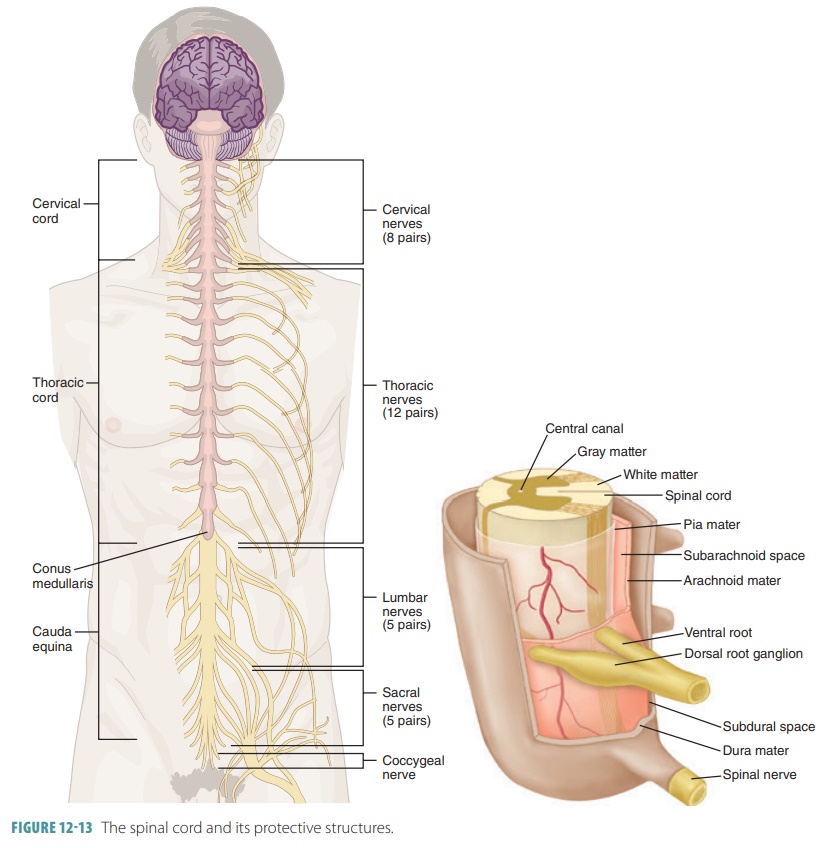
Pages 3 Ratings 100 19 19 out of 19 people found this document helpful. The spinal cord extends caudally from the brainstem running from the medullary-spinal junction at about the level of the first cervical vertebra to about the level of the twelfth thoracic vertebra see Figure 110The vertebral column and the spinal cord within it is divided into cervical thoracic lumbar sacral and coccygeal regions. The spinal cord is made of gray and white matter just like other parts of the CNS.
Progressive wear-and-tear of the spinal disks in your neck cervical disks that can press on the spinal cord cervical. The spine is made up of vertebrae back bones that protect and surround the spinal cord which is a column of nerve tissue. Describe the structure of a spinal nerve.
The spinal nerve emerges from the spinal column through an opening intervertebral foramen between adjacent vertebrae. Each spinal nerve emerges from the spine through the foramen. Inflammation in one or more segments of your spinal cord.
These nerves are attached to the spinal cord by two roots- dorsal sensory root and ventral motor root. Describe the structure of a spinal nerve Spinal nerve is part of the PNS somatic. The dorsal root is also called the posterior or sensory root.
Spinal nerves function. The 31 pairs of spinal nerves are as follows. These two roots unite and form a spinal nerve which extends through the intervertebral foramen.
The spine is made up of a column of vertebral bones that surround and protect the spinal cord. Course Title BIOL 241. A spinal nerve emerges at two points from the spinal cord the ventral and dorsal roots.
Spinal Nerves. Common conditions that can affect your spinal cord include. The spinal nerves are present on each side of the spine within a few centimeters.
This is true for all spinal nerves except for the first spinal nerve. Eight pairs of cervical spinal nerves C1-C8 Twelve pairs of thoracic spinal nerves T1-T12 Five pairs of lumbar spinal nerves L1-L5 Five pairs of sacral spinal nerves S1-S5. Spinal nerve The nerve that occurs at the joining of the dorsal and ventral roots dorsal primary ramus The first posterior branch of the spinal nerve ventral primary ramus The first anterior branch of the spinal nerve efferent A nerve that leaves the central nervous system motor afferent A nerve that enters the central nervous system sensory.
The top part of the spine has seven vertebrae C1 to C7. These are openings on the left and right sides of the vertebral bones of the spine.

Human Spine Drawing Art Human Spine Spine Drawing Anatomy Drawing

Spinal Nerve Definition Function Diagram Number Facts Britannica
Spinal Cord Central Nervous System
Peripheral Nervous System The 31 Pairs Of Spinal Nerves Exit The Spinal Cord To The Right And Left Sides Peripheral Nervous System Nervous System Spinal Nerve


0 Comments